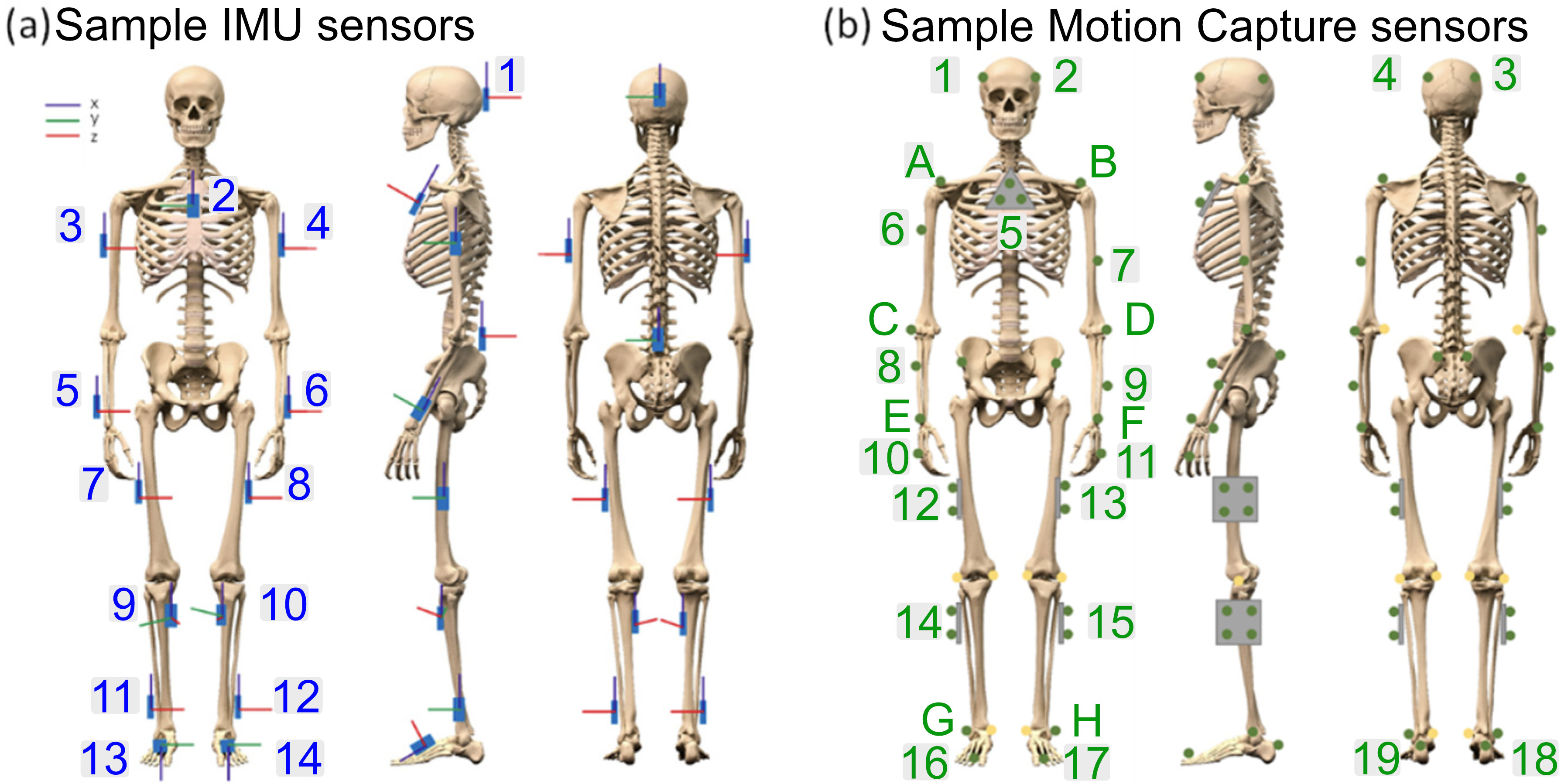
Anatomical Table

| Hanavan Body Part | HED Label | HED Description | Anatomical Description | Fiducials | Coordinate System | Example IMU | Example Mocap | Landmark visualization | |
|---|---|---|---|---|---|---|---|---|---|
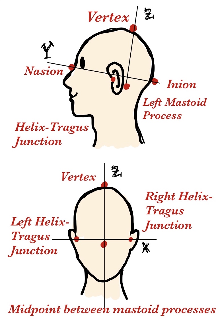
| 1 | Head | The upper part of the human body, or the front or upper part of the body of an animal, typically separated from the rest of the body by a neck, and containing the brain, mouth, and sense organs. | The structure superior to the neck, encasing the brain, eyes, ears, nose, and mouth. It is demarcated by the skull, which separates the head from the neck at the base of the skull. | Nasion Inion Left Helix-Tragus Junction (LHJ) Right Helix-Tragus Junction(RHJ) Vertex Midpoint between Mastoid processs(MMP) |
X: LHJ → RHJ Y: Inion → Nasion Z: MMP → Vertex |
1. (50, 0, 50) | 1. (100, 100, 80) 2. (0, 100, 80) 3. (100, 10, 80) 4. (0, 10, 80) |
 |
|
| n/a | Neck | The part of the body connecting the head to the torso, containing the cervical spine and vital pathways of nerves, blood vessels, and the airway. | The anatomical region bounded by the base of the skull, and the clavicles inferiorly, containing vital structures such as the cervical vertebrae, spinal cord, carotid arteries, and trachea. | C7 vertebra (C7) Suprasternal Notch (Jugular Notch) Left Mastoid process(LMP) Right Mastoid process (RMP) Midpoint between Mastoid processs(MMP) |
X: LMP → RMP Y: C7 → Jugular Notch Z: C7 → MMP |
n/a | n/a | 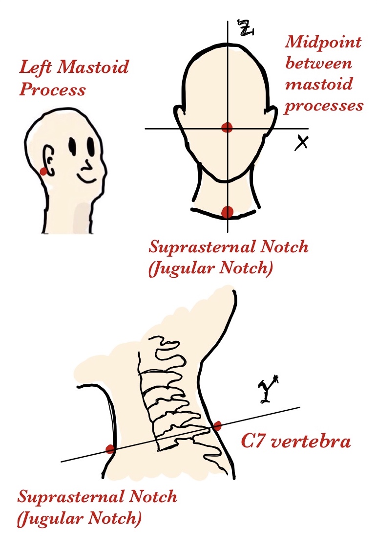 |
|
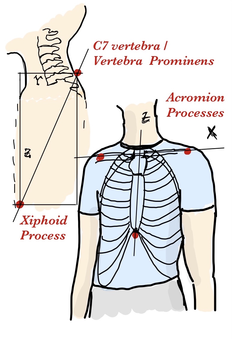
| 2 | Torso-chest | The body excluding the head and neck and limbs. | The upper part of the torso, extending from the base of the neck to the diaphragm. It’s framed by the rib cage, which includes the ribs, sternum, and thoracic vertebrae. The thorax houses vital organs such as the heart and lungs. | Left Acromion Process (LAP) Right Acromion Process(RAP) C7 vertebra (C7) Xiphoid Process |
X: LAP → RAP Y: C7 → Xiphoid Process (shorter axis) Z: C7 → Xiphoid Process (longer axis) |
2. (50, 100, 70) | 5. (50, 100, 70) A.(100, 100, 30) B.(0, 100, 30) |
 |
|
| 3 | Abdomen | The body excluding the head and neck and limbs. | The lower part of the torso, situated between the thorax and the pelvis. It is bounded superiorly by the diaphragm and inferiorly by the pelvic girdle. The abdomen contains important digestive, urinary, and reproductive organs, and is supported by various abdominal muscles. | Umbilicus (Navel) Left Anterior Superior Iliac Spine (Left ASIS) Right Anterior Superior Iliac Spine (Right ASIS) Posterior Superior Iliac Spine (PSIS) Xiphoid Process |
X: Left ASIS → Right ASIS Y: midpoint ASIS → midpoint PSIS Z: Navel → Xiphoid Process |
n/a | n/a | 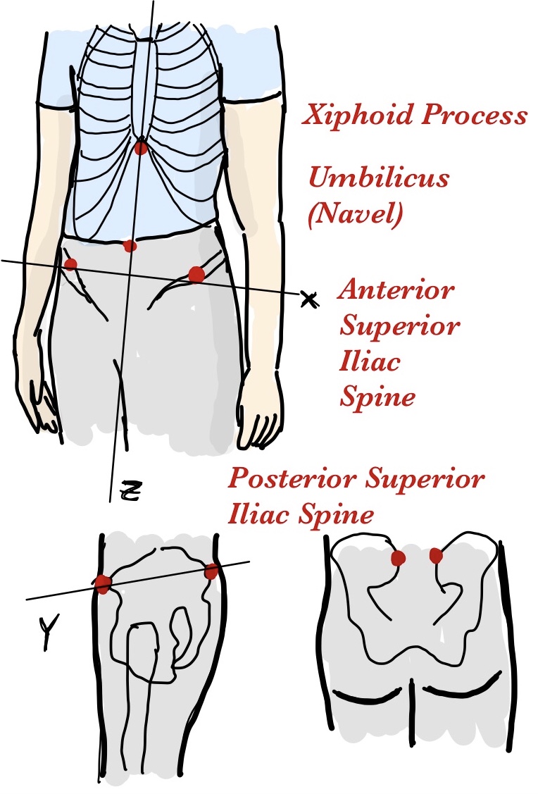 |
|
| 4 | (Right, Hand) | The distal portion of the upper extremity. It consists of the carpus, metacarpus, and digits. | The terminal part of the forearm, beginning at the wrist joint (where the radius and ulna meet the carpal bones) and extending to the fingertips, including the carpus, metacarpus, and phalanges. | Radial Styloid Process (RSP) Ulnar Styloid Process (USP) Third Metacarpophalangeal Joint (MCP): The knuckle of the middle finger |
X: RSP → USP Y: Dorsal → Palmar surface at the midpoint RSP-USP Z: MCP → midpoint RSP-USP |
n/a | 10. (100, 50, 50) | 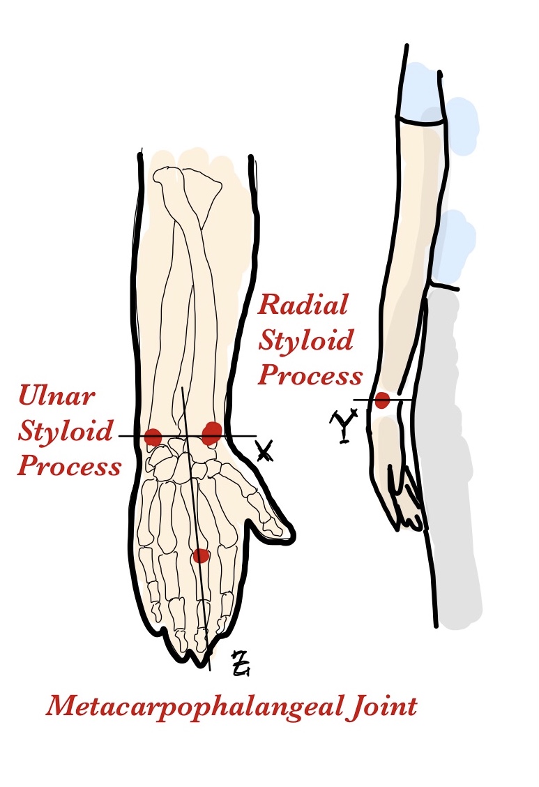 |
|
| 5 | (Left, Hand) | The distal portion of the upper extremity. It consists of the carpus, metacarpus, and digits. | The terminal part of the forearm, beginning at the wrist joint (where the radius and ulna meet the carpal bones) and extending to the fingertips, including the carpus, metacarpus, and phalanges. | Radial Styloid Process (RSP) Ulnar Styloid Process (USP) Third Metacarpophalangeal Joint (MCP): The knuckle of the middle finger |
X: USP → RSP Y: Dorsal → Palmar surface at the midpoint RSP-USP Z: MCP → midpoint RSP-USP |
n/a | 11. (0, 50, 50) | 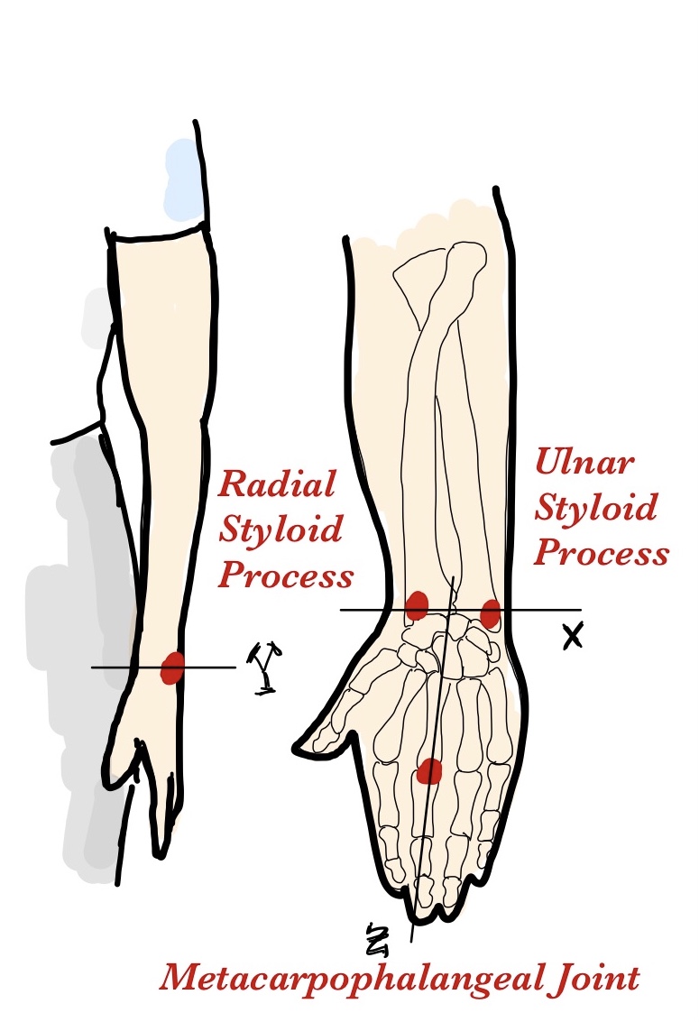 |
|
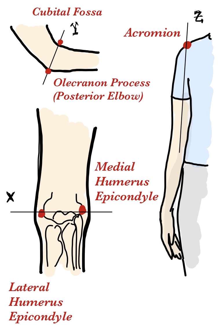
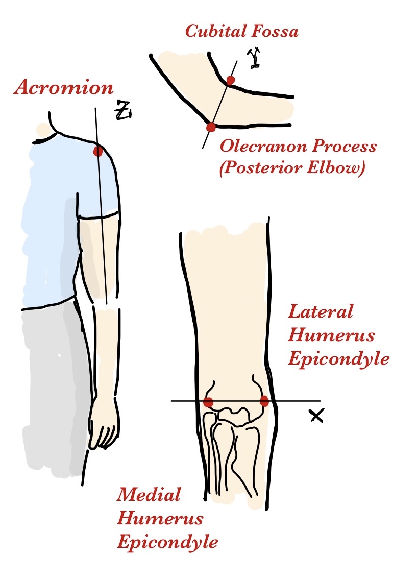
| 6 | (Right, Upper-arm) | Portion of arm between shoulder and elbow. | The segment between the shoulder and elbow, outlined by the humerus bone. It starts at the glenohumeral joint and ends at the elbow joint where the humerus meets the radius and ulna. | Acromion Medial Humerus Epicondyle (MHE) Lateral Humerus Epicondyle (LHE) Posterior Elbow (Olecranon Process) Cubital Fossa |
X: MHE → LHE Y: Olecranon Process → Cubital Fossa Z: midpoint MHE-LHE → Acromion |
3. (100, 50, 70) | 6. (100, 50, 70) C. (100, 50, 0) |
 |
|
| 7 | (Left, Upper-arm) | Portion of arm between shoulder and elbow. | The segment between the shoulder and elbow, outlined by the humerus bone. It starts at the glenohumeral joint and ends at the elbow joint where the humerus meets the radius and ulna. | Acromion Medial Humerus Epicondyle (MHE) Lateral Humerus Epicondyle (LHE) Posterior Elbow (Olecranon Process) Cubital Fossa |
X: MHE → LHE Y: Olecranon Process → Cubital Fossa Z: midpoint MHE-LHE → Acromion |
4. (0, 50, 70) | 7. (0, 50, 50) D. (0, 50, 0) |
 |
|
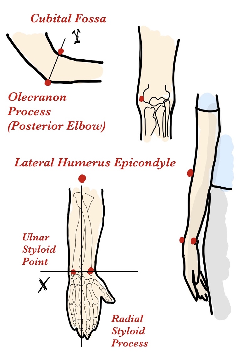
| 8 | (Right, Forearm) | Lower part of the arm between the elbow and wrist. | Extends from the elbow to the wrist, demarcated by the radius and ulna. Begins at the elbow joint and ends at the wrist joint. | Radial Styloid Process (RSP) Ulnar Styloid Process (USP) Lateral Humerus Epicondyle (LHE) Posterior Elbow (Olecranon Process) Cubital Fossa |
X: RSP → USP Y: Right-hand rule (limits: Olecranon Process → Cubital Fossa) Z: midpoint RSP-USP → LHE |
5. (100, 50, 30) | 8. (100, 50, 70) E. (50, 100, 0) |
 |
|
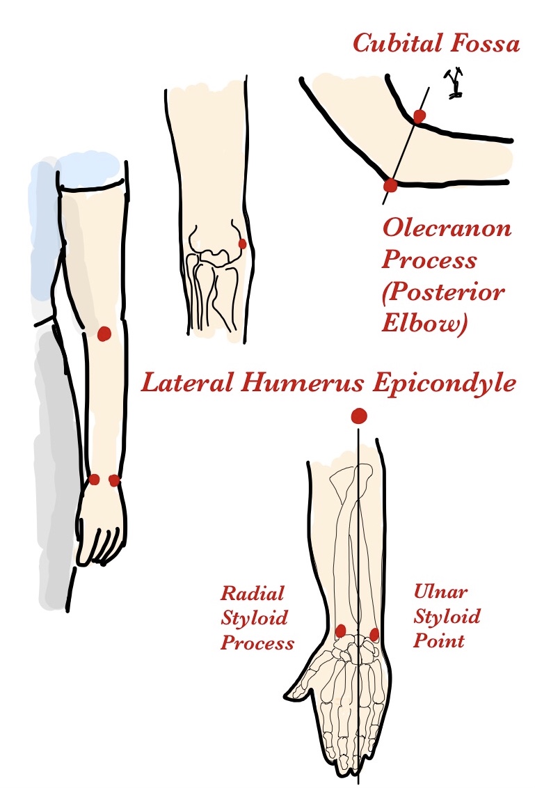
| 9 | (Left, Forearm) | Lower part of the arm between the elbow and wrist. | Extends from the elbow to the wrist, demarcated by the radius and ulna. Begins at the elbow joint and ends at the wrist joint. | Radial Styloid Process (RSP) Ulnar Styloid Process (USP) Lateral Humerus Epicondyle (LHE) Posterior Elbow (Olecranon Process) Cubital Fossa |
X: USP → RSP Y: Right-hand rule (limits: Olecranon Process → Cubital Fossa) Z: midpoint RSP-USP → LHE |
6. (0, 50, 30) | 9. (0, 50, 50) E. (50, 100, 0) |
 |
|
| 10 | (Right, Thigh) | Upper part of the leg between hip and knee. | The part of the lower limb extending from the hip to the knee, primarily composed of the femur. It starts at the hip joint (where the femur connects with the pelvic girdle) and ends at the knee joint. | Greater Trochanter Femur Medial Epicondyle (FME) Femur Lateral Epicondyle (FLE) Patellar Anterior Surface Back of the knee (Popliteal Fossa) |
X: FME → FLE Y: Popliteal Fossa → Patellar Anterior Surface Z: midpoint FME-FLE → Greater Trochanter |
7. (100, 50, 50) | 12. (100, 50, 50) | 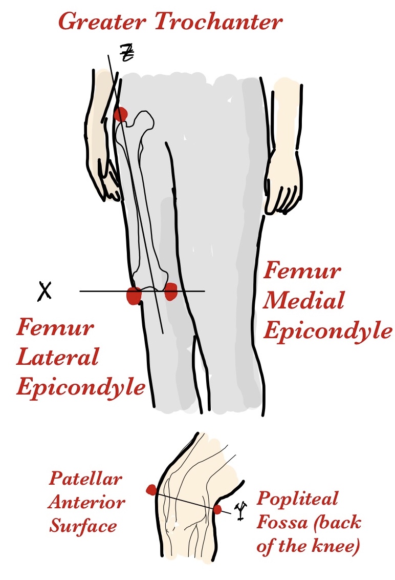 |
|
| 11 | (Left, Thigh) | Upper part of the leg between hip and knee. | The part of the lower limb extending from the hip to the knee, primarily composed of the femur. It starts at the hip joint (where the femur connects with the pelvic girdle) and ends at the knee joint. | Greater Trochanter Femur Medial Epicondyle (FME) Femur Lateral Epicondyle (FLE) Patellar Anterior Surface Back of the knee (Popliteal Fossa) |
X: FLE → FME Y: Popliteal Fossa → Patellar Anterior Surface Z: midpoint FME-FLE → Greater Trochanter |
8. (0, 50, 50) | 13. (0, 50, 50) | 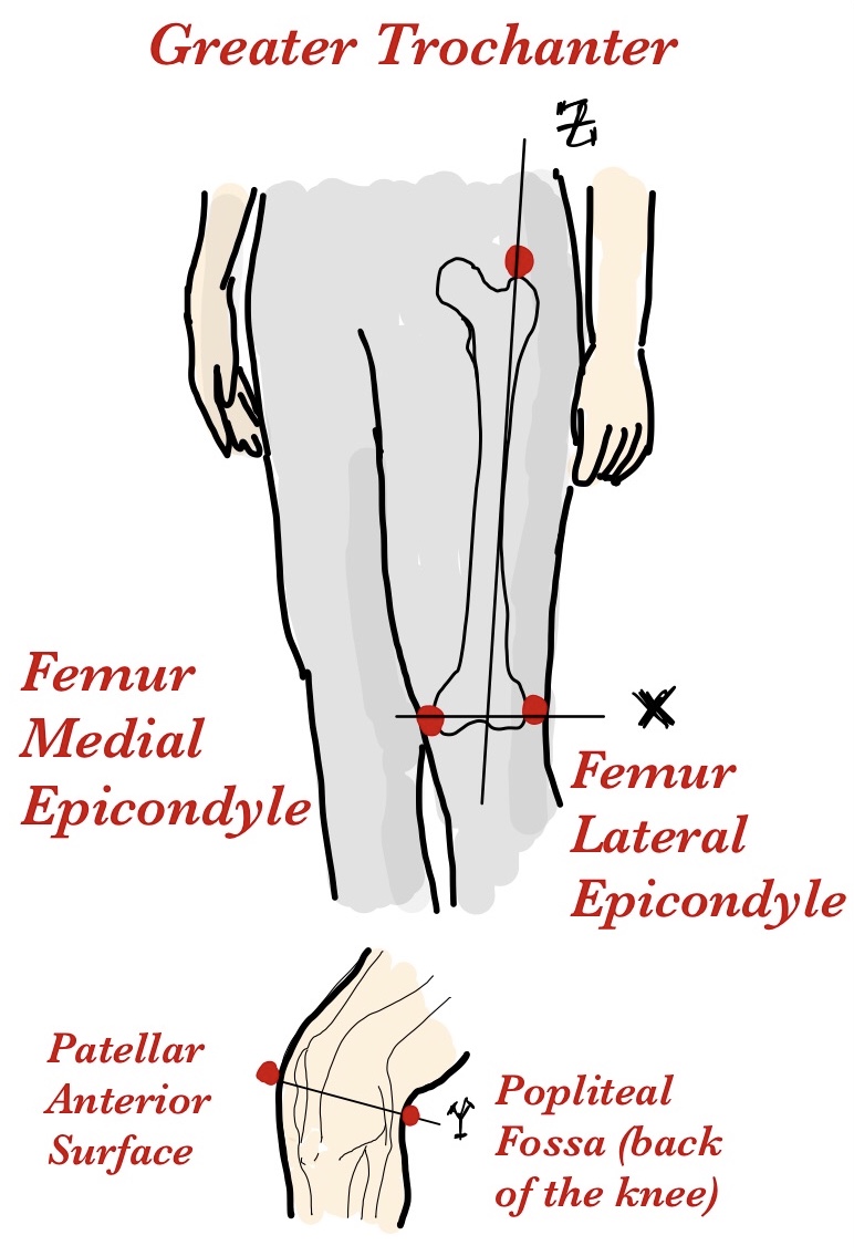 |
|
| 12 | (Right, Lower-leg) | The part of the leg between the knee and the ankle. | Calf: The posterior segment of the lower leg, from the knee to the ankle. The calf muscles (gastrocnemius and soleus) overlay the tibia and fibula, starting below the knee joint and extending to the ankle. Shin: The anterior part of the lower leg, starting just below the knee joint and extending to the ankle. It primarily involves the tibia. | Lateral Malleolus (LM) Medial Malleolus (MM) Patellar Anterior Surface Back of the knee (Popliteal Fossa) |
X: MM → LM Y: Popliteal Fossa → Patellar Anterior Surface Z: midpoint MM-LM → Patellar Anterior Surface |
9. (0, 50, 80) 11. (100, 50, 20) |
14. (100, 50, 80) G. (100, 50, 0) |
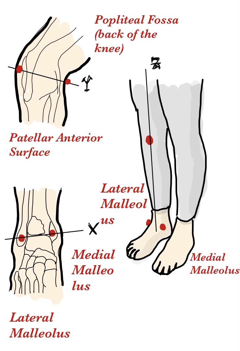 |
|
| 13 | (Left, Lower-leg) | The part of the leg between the knee and the ankle. | Calf: The posterior segment of the lower leg, from the knee to the ankle. The calf muscles (gastrocnemius and soleus) overlay the tibia and fibula, starting below the knee joint and extending to the ankle. Shin: The anterior part of the lower leg, starting just below the knee joint and extending to the ankle. It primarily involves the tibia. | Lateral Malleolus (LM) Medial Malleolus (MM) Patellar Anterior Surface Back of the knee (Popliteal Fossa) |
X: LM → MM Y: Popliteal Fossa → Patellar Anterior Surface Z: midpoint MM-LM → Patellar Anterior Surface |
10. (100, 50, 80) 12. (0, 50, 20) |
15. (0, 50, 80) G. (0, 50, 0) |
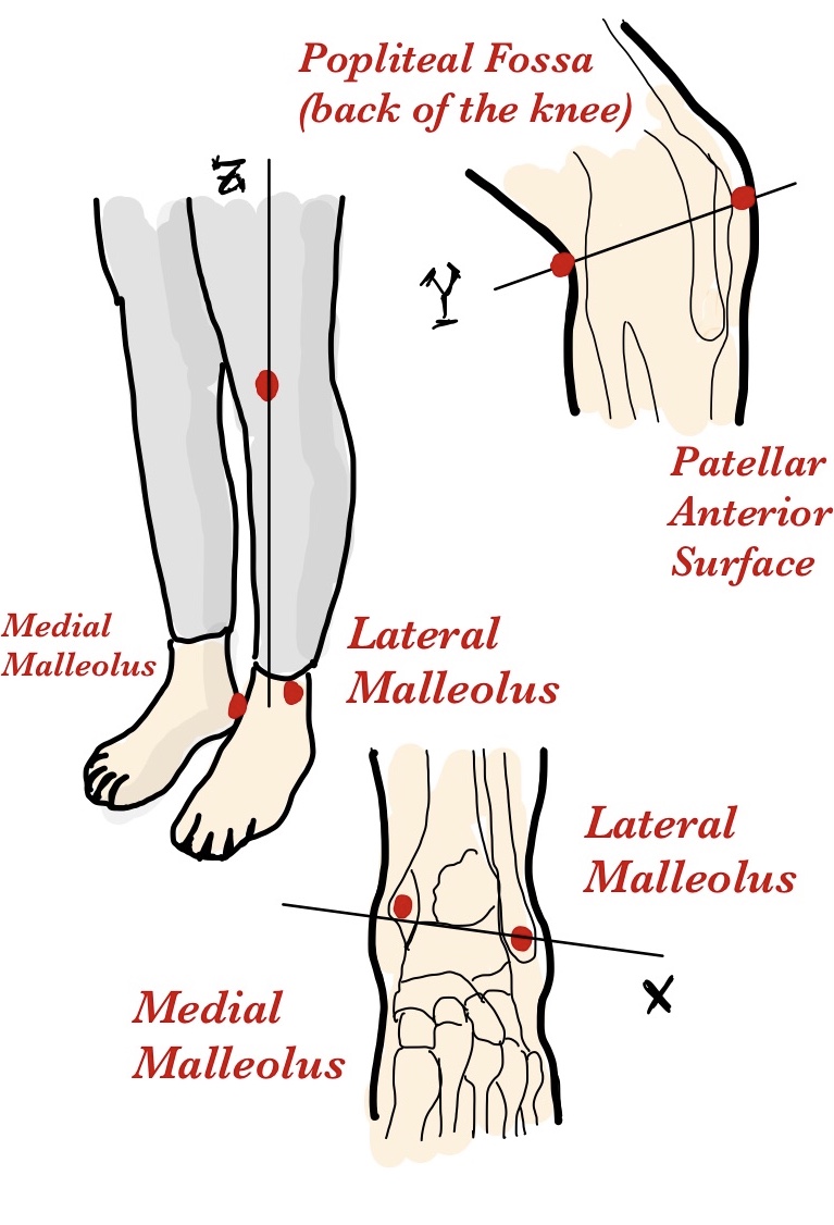 |
|
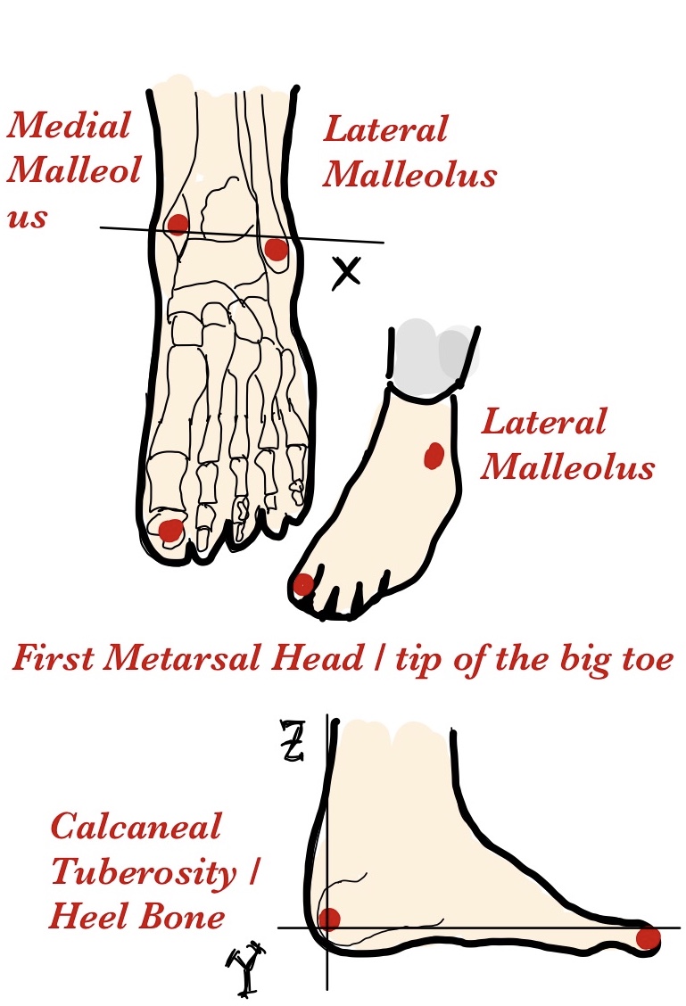
| 14 | (Right, Foot) | The structure found below the ankle joint required for locomotion. | The distal extremity of the lower leg, starting at the ankle joint (where the tibia, fibula, and talus meet) and extending to the tips of the toes. It includes the tarsus, metatarsus, and phalanges. | Heel Bone, Calcaneal Tuberosity (CT) Tip of the big toe, First Metatarsal Head (FME) Lateral Malleolus (LM) Medial Malleolus (MM) |
X: MM → LM Y: CT → FME Z: CT to MM-LM line |
13. (50, 60, 80) | 16. (50, 100, 40) 18. (50, 0, 50) |
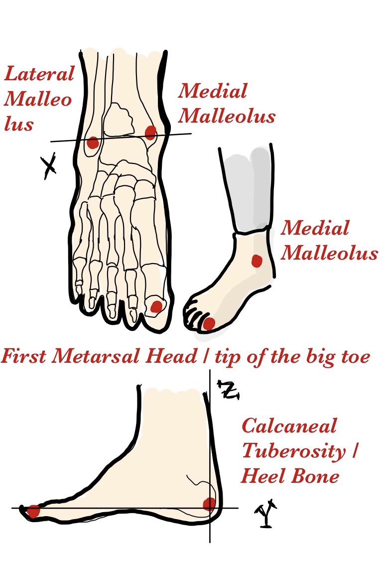 |
|
| 15 | (Left, Foot) | The structure found below the ankle joint required for locomotion. | The distal extremity of the lower leg, starting at the ankle joint (where the tibia, fibula, and talus meet) and extending to the tips of the toes. It includes the tarsus, metatarsus, and phalanges. | Heel Bone, Calcaneal Tuberosity (CT) Tip of the big toe, First Metatarsal Head (FME) Lateral Malleolus (LM) Medial Malleolus (MM) |
X: LM → MM Y: CT → FME Z: CT to MM-LM line |
14. (50, 60, 80) | 17. (50, 100, 40) 19. (50, 0, 50) |
 |
NOTE: Joint markers should be annotated in the same coordinate system as their proximal body part. For example, the right shoulder joint marker should be annotated in the same coordinate system as torso, and the right elbow joint marker should be annotated in the same coordinate system as the right upper-arm.
